
We aims to develop rapid molecular diagnostic methods as well as understand the pathogenesis of ocular infections and host pathogen interactions. It employs various conventional microbiological methods and molecular tools such as conventional PCR, Real Time PCR, DNA barcoding and Next generation sequencing. Conventional and Real Time PCR are routinely employed for the identification and quantification of known ocular pathogens including bacteria, fungus, Acanthamoeba spp., Mycobacterium spp., Toxoplasma, Microsporidia and virus such as HSV, VZV and CMV. For identification of unknown bacterial and fungal pathogens, 16S and 28S rDNA gene barcoding by sequencing are employed respectively. Major ocular pathogens currently under study include methicillin resistant Staphylococcus aureus (MRSA), extended spectrum β-lactamase producers (ESBL), Pseudomonas aeruginosa, Nocardia spp., Acanthamoeba spp. and Trematodes. The department also has a large repository of ocular isolates which enables the team to probe into the virulence and antibiotic resistance mechanisms of pathogens in the context of clinical treatment. Multi locus sequence analysis is employed for the identification of the closely related pathogens such as Fusarium spp., Nocardia spp., and Acanthamoeba genotypes. The host pathogen interactions are studied using exvivo and invitro models of ocular infection.
Research Areas
Bacterial Keratitis
Infectious keratitis is a major cause of blindness, next to cataract and majority of cases are caused by either fungi or bacteria in developing countries. Bacterial keratitis develops very quickly because of its rapid progression and increased corneal destruction and it may lead to blindness, if left untreated. At the microbiology lab, ocular isolates of P. aeruginosa and methicillin resistant S. aureus (MRSA) are used to delineate their virulence and antibiotic resistance mechanisms. The pathogenesis of P. aeruginosa is mediated through the production of several cell-associated and extracellular virulence factors. The department studies the involvement of Type 3 secretion system and other virulence factors of P. aeruginosa in causing ocular infection by both phenotypic and genotypic methods. Analysis of the differentially expressed genes by whole genome sequencing and transcriptome analysis of the ocular isolates is also being done. Another study focuses on the molecular characterization and epidemiological analysis of MRSA causing ocular infections. The team found that community acquired MRSA strains (SCCmec type IV and V) are more prevalent than hospital acquired MRSA in causing ocular infections. Further studies are done to analyse the expression of toxin genes in association with various ocular infections using advanced molecular methods.
Host-Pathogen Interactions
Since corneal ulceration is a leading cause of ocular morbidity and blindness worldwide, it is imperative that the functional and molecular aspects of the disease be well understood to develop an effective treatment plan. An earlier study from the lab reported elevated expression of TLRs (Toll Like Receptors) in relation to Microbial keratitis (Karthikeyan et al., 2013). Although much of the focus in the literature has been on the study of protein regulators of inflammation, miRNAs have emerged as important controllers of certain features of the inflammatory process. The team proposed that miRNAs may play a role in the pathogenesis of microbial keratitis by modulating TLR signaling which has not been previously reported. The department has undertaken a study to reveal the involvement of miRNAs in microbial keratitis pathogenesis by expression profiling of miRNAs in corneal scrapings from keratitis and normal controls. For an effective therapeutic intervention of keratitis without the need for surgery, excessive inflammatory responses need to be curtailed. Involvement of various host innate defense molecules in ocular inflammation caused by infectious keratitis is being analysed.
Ocular Parasitic Infections
Pediatric parasitic ocular inflammation is one of the common clinical conditions in Southern India. The department has identified the causative agent of granulomatous uveitis as trematode, Procerovum varium and source of infection as fresh water snails, Melanoid tuberculata. Immunohistochemistry studies are now being done to analyze the inflammatory cellular pattern to understand the disease pathogenesis. Another vision threatening ocular infection is caused by Acanthamoeba which leads to corneal ulceration, loss of visual acuity, corneal scarring and monocular blindness. Rate of incidence of Acanthamoeba was 3.1% in 2013, at Aravind Eye Hospital, Madurai, India. Acanthamoeba keratitis is difficult to treat and there is minimal evidence on which to base treatment decisions. The department is investigating the microbiological clearance time for Acanthamoeba by Real time quantitative PCR. Also, Minimum Cysticidal Concentration (MCC) of anti-microbial drugs is being determined with a view to evaluate the efficacy of combination therapy by invitro synergy assay.
Ongoing Projects
- Quantification of the Acanthamoeba by Real Time PCR in keratitis patients.
- Nocardia species identification by Mutli locus sequence analysis (MLSA)
- Characterization of the virulence determinants of pseudomonas aeruginosa causing ocular infections.
- Genotypic characterization and analysis of virulence factors in Methicillin resistant staphylococcus aureus causing ocular infections.
- Microbiological clearance time and Sensitivity assay for acanthamoeba keratitis
- Asian Corneal Society for Infectious Keratitis study (ACSIKS)- A multicentric prospective, clinical study on fungal keratitis presenting in eight countries (India, China, Japan, South Korea, Taiwan, Thailand, Philippines, and Singapore).
- Diagnostic Markers for Ocular Tuberculosis, funded by DBT, India
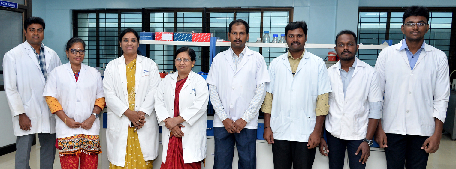
Faculty
- Dr. Lalitha Prajna, Clinician – Research Scientist
- Dr. S.R. Rathinam, Clinician – Research Scientist
- Dr. D. Bharanidharan, Scientist
Research Associate
- Mr. R. SivaGanesa Karthikeyan
Technician
- Mr. A. Selvapandiyan
PCR Machines
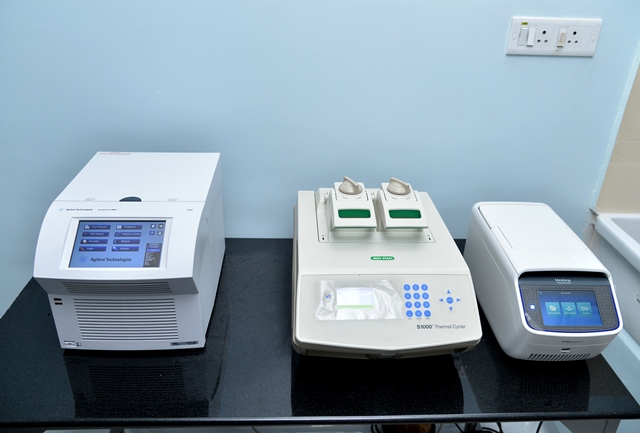
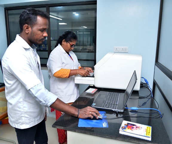
Microbiology lab has a separate facility for PCR machine and has four conventional and one gradient PCR machine. All these machines are used routinely for molecular diagnosis of ocular pathogens.
Molecular Devices - M3 Spectra Max Multimode Reader
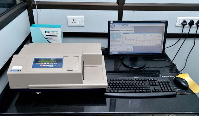
Multimode reader is equipped with visible and UV light used for bacterial growth curve analysis, ELISA and protein estimation procedure. Samples can be analyzed in 96 well micro plates or cuvettes.
ABI Quantstudio 5 Real Time PCR
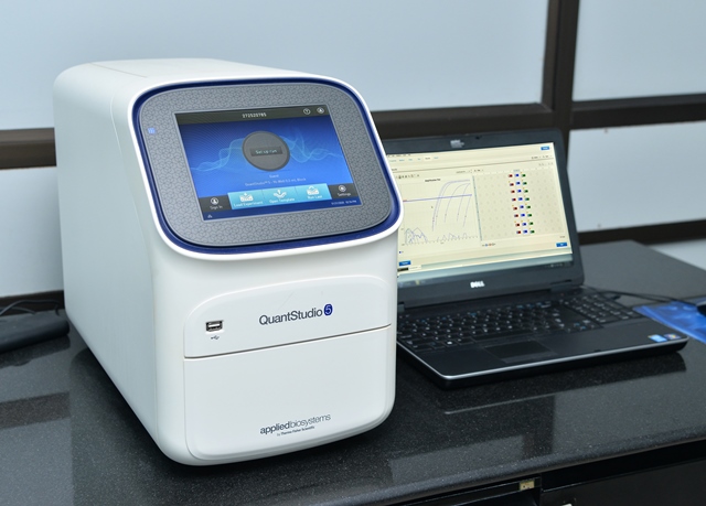
Real Time PCR is mainly used for diagnosis of ocular pathogens such as bacteria, fungus, Acanthamoeba, Toxoplasma, Mycobacterium tuberculosis and virus (HSV, VZV and CMV).Other departments use this machine to study gene expression, deletion/duplication analysis and copy number variations.