
We aim to understand the pathogenesis of eye diseases and take forward the translational leads from the basic research to develop diagnostic or prognostic methods that can help in improving the clinical management of the disease in patients. We have been working on major eye diseases namely, fungal keratitis, diabetic retinopathy, glaucoma, and keratoconus.
Research projects at the Proteomics Department are supported through funds from Government agencies including DBT, DHR and SERB as well as private organisations including Mind Tree Organization, Cognizant Foundation and Sun Pharma. We also collaborate with international research groups that includes Indo-French collaboration with Institut Pasteur, France for fungal keratitis, Indo-UK collaboration with Moorefields Hospital, London for diabetic retinopathy, and Indo-US collaboration for primary open angle glaucoma.
Fungal Keratitis
Our group is perhaps the largest group working on fungal infection of the eye in India as well as across the globe. Fusarium and Aspergillus are the major fungal pathogens causing fungal keratitis in India. Agricultural workers are the major group affected by this infection primarily due to their frequent exposure to fungal spores and accidental trauma to the eye.
We adopted a comprehensive OMICS approach to decipher the pathogenesis of mycotic keratitis, at the level of both the human host and the fungal pathogens. On the host side, proteomics of tear and cornea helped unravel the major events/pathways activated in the human host in response to the fungal infection. We have also identified many proteins in the tear fluid as indicators of the severity of the infection. We are currently validating many of these tear markers to develop a method to predict the prognosis of the fungal ulcer. Genomics, transcriptomics and proteomics of the fungal pathogens has led to a comprehensive understanding on inter- as well as intra-species difference among the fungal pathogens.
In addition, through a collaborative study with the Institut Pasteur, Paris we have examined in detail the two Aspergillus pathogens, A.flavus (a predominant corneal pathogen) and A. fumigautus (primary lung pathogen). This study compared the cell wall architecture of both these pathogens and how they influence the host innate immune response.
Diabetic Retinopathy
Diabetic retinopathy (DR) is one of the most common microvascular complication in diabetic and is a leading cause of vision loss among working-age adults in the world. The clinical classification of DR is graded into two major groups, non-proliferative diabetic retinopathy (NPDR) and proliferative diabetic retinopathy (PDR). The main cause of visual impairment in DR is the development of diabetic macular edema (DME) and PDR.
Our research focus on DR is to identifyDR-specific, protein biomarkers. Towards this, we have examined the proteome of both serum and vitreous to identify proteins that show significant alteration with reference to the disease stage and/or disease progression. In addition to the serum proteins, we also analyse the proteomeof microparticles (vesicles secreted by different cell types in the body) circulating in the serum for identification and validation of DM and DR-specific protein markers. Through an Indo-UK collaborative project, we are currently validating a panel of selected serum markers in both Indian and UK population. Through these biomarkers studies, we aim to identify diagnostic markers that will help identify DM patients at high risk of developing DR as well as prognostic markers that will predict the disease progression among DR patients.
Glaucoma
Primary Open Angle Glaucoma (POAG) is the second leading cause of blindness worldwide, contributing to 12% of world blindness. POAG is asymptomatic and the loss of vision is irreversible. Proteome analysis of aqueous humorfrom POAG patients in comparison with that of cataract control enabled the identification of POAG related protein changes. These proteins will be validated in a larger patient cohort to develop a panel for diagnostic or prognostic use.
Pseudoexfoliation glaucoma is yet another glaucomatous condition that is quite prevalentand nearly 25-35% of the PEXS patients develop glaucoma in India. There is no treatment available for PEXS as the source and mechanism of formation of PEX material is currently unclear. To address this gap in knowledge, we are examining the role of iris tissue in the pathogenesis of PEX disease.
Ongoing Projects
- Project title: Proteome profiling of serum microparticles in diabetes and diabetic retinopathy patients: Towards identification and validation of predictive biomarkers – Funding: Department of Health Research, Govt of India
- Project title: Prospective Multicenter discovery and validation of diagnostic circulating and urinary biomarkers and development of sensor(s) to detect sight threatening diabetic retinopathy: Biomarker and Biosensor study in UK and India (Work Package 3) – Funding: Research Councils UK
- Project title: Understanding the role of iris in pseudoexfoliation disease – Funding: Aravind Medical Research Foundation
- Project title: Prediction of Treatment Outcome in Fungal Keratitis Patients (to be initiated) – Funding: Cognizant Foundation
- Project title: Development of aptamer-based assays for diagnosis of infectious keratitis and absolute quantitation of proteoform markers of diabetic retinopathy (to be initiated) – Funding: Sun Pharma
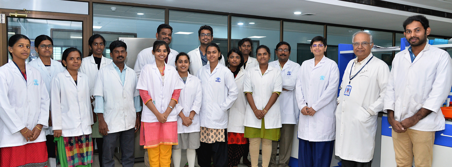
Faculty
- Prof. K. Dharmalingam, Director – Research
- Dr. Jeya Maheswari, Scientist
- Dr. O.G. Ram Prasad, Scientist
Clinician Scientist
Collaborators
- Prof. Colin E Willoughby, University of Liverpool, UK
- Prof. Jean-Paul Latge, Institut Pasteur, France
- Prof. Shoba Sivaprasad, Moorefields Hospital, London
Research Scholars
- A. Divya
- P. Vignesh
- Irene Daniel
- Guhan
- T.M. Nasrin Banu
- G. Hariharan
- K.R. Shruthi Mahalakshmi
- R. Mathumathi
- Nishanthi Sudhakar
- Dr. Sagnik Sen (Research Fellow-Retina)
Lab Assistants
- T.Parameshwari
- M.Sujitha
- M.Manikandan
- M.Dhanalakshmi
- T.Maheswari
- K.PackiyaLakshmi
- V.Renuka
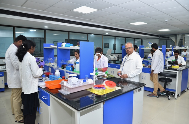
We have a state-of-the-art infrastructure for carrying out proteomics research that includes the following facilities:
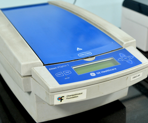
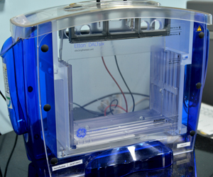
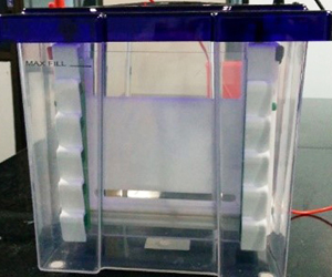
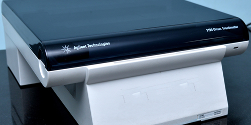
1D and 2D facilities
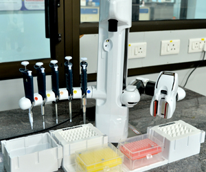
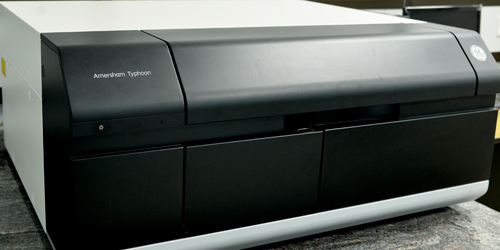
A self-sustained lab with all the facilities needed for carrying out proteomics research using different approaches such spectrophotometer, centrifuges, 1D SDS-PAGE, IEF focussing unit as well as fractionator, 2D polyacrylamide electrophoresis units, and for quantitative proteome analysis using fluorescent labels (DIGE laser scanner). In addition we also have Andrew liquid handling robot for highthroughput handling of samples for proteomics as well as qPCR experiments.
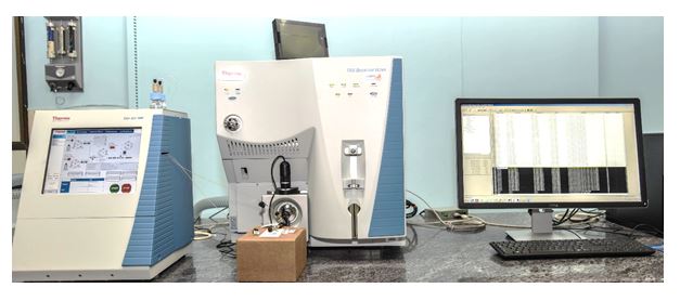
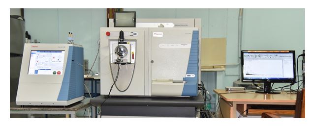
Mass spectrometry facility
The core of the proteomics facility is the availability of two mass spectrometers
- nanoLC-Orbitrap velos pro mass spectrometer: This is a high-resolution mass spectrometer that enables the in-depth identification and quantitative comparison of proteins in a complex proteome such as tear, serum, corneal tissue, fungal cells etc.
- uHPLC/nanoLC-Triple Stage Quadrupole mass spectrometer: This mass spectrometer is used for a high-throughput, reproducible, and targeted quantitation of proteins in complex proteome
Other facilities
- Cell culture lab
- Fungal lab
- Clean room for RNA and qPCR studies